Retinal Detachment

Retinal Detachment

Retinal detachment is a serious eye condition in which the retina, the thin layer of tissue at the back of the eye responsible for capturing and transmitting visual signals to the brain, becomes separated from its normal position. This separation can lead to a loss of vision if not promptly treated.

Symptoms

If you have Retinal Detachment you may observe

...

Flashing Lights

...

Shadow or Curtain Effect

...

Blurred Vision

...

Loss of Vision

Vision screenings using adequate testing equipment.

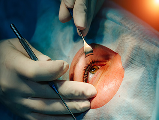
Retinal detachment requires immediate medical attention. Treatment options typically involve surgical intervention to reattach the retina. Different procedures, such as pneumatic retinopexy, scleral buckle, or vitrectomy, may be utilized depending on the severity and location of the detachment.

Prompt diagnosis and treatment are crucial in preserving vision. Regular comprehensive eye exams can help detect early signs of retinal detachment, especially for individuals at higher risk due to specific eye conditions or previous trauma. If any symptoms of retinal detachment arise, seeking immediate medical attention is essential to prevent permanent vision loss.

Treatments for Retinal Detachment

Injecting air or gas into your eye In this procedure, called pneumatic retinopexy (RET-ih-no-pek-see), the surgeon injects a bubble of air or gas into the center part of the eye (the vitreous cavity). If positioned properly, the bubble pushes the area of the retina containing the hole or holes against the wall of the eye, stopping the flow of fluid into the space behind the retina.

Indenting the surface of your eye. This procedure, called scleral (SKLAIR-ul) buckling, involves the surgeon sewing (suturing) a piece of silicone material to the white of your eye (sclera) over the affected area. This procedure indents the wall of the eye and relieves some of the force caused by the vitreous tugging on the retina.

Draining and replacing the fluid in the eye In this procedure, called vitrectomy (vih-TREK-tuh-me), the surgeon removes the vitreous along with any tissue that is tugging on the retina. Air, gas or silicone oil is then injected into the vitreous space to help flatten the retina.

FAQ-Retinal Detachment Surgery at Sarvdrishti Hospital

Detachment of retina at the macular area is the cause for loss of vision.

Retinal detachment can occur due to various factors, such as age-related changes in the vitreous gel, trauma or injury to the eye, underlying eye conditions like lattice degeneration or retinoschisis, and certain medical conditions like diabetic retinopathy.

Several risk factors can increase the likelihood of developing retinal detachment, including being over the age of 40, having a family history of retinal detachment, being nearsighted (myopia), previous eye surgeries or procedures, and experiencing eye trauma or injury.

Yes, retinal detachment is considered a medical emergency. Prompt medical attention is crucial because delays in treatment can lead to permanent vision loss.

While it may not be possible to prevent all cases of retinal detachment, certain measures can help reduce the risk.